Exploring the Role of Kinetic Chain Dysfunction in Orthopedic Injuries

Exploring the Role of Kinetic Chain Dysfunction in Orthopedic Injuries

Explore the role of kinetic chain dysfunction in orthopedic injuries and understand how imbalances can lead to a variety of musculoskeletal problems. This comprehensive overview highlights key factors contributing to these injuries and offers insights for prevention and management.

How does proximal stability influence distal mobility in the context of kinetic chain dysfunction and its associated orthopedic injuries?

Proximal stability plays a crucial role in facilitating distal mobility, particularly when considering the kinetic chain and its dysfunctions that often lead to orthopedic injuries. In the human body, proximal joints such as those in the pelvis and spine provide essential support for more distal segments like the knees, ankles, shoulders, and wrists. When proximal stabilization is compromised due to factors like muscle weakness or poor posture, it can result in compensatory movement patterns that increase stress on distal joints during dynamic activities—this phenomenon is commonly associated with conditions such as overuse injuries or repetitive strain syndromes. For instance, if core muscles fail to stabilize effectively during sports movements like running or throwing, this lack of stability may force adjacent structures into abnormal ranges of motion which could ultimately cause joint pain or even tears in ligaments and tendons downstream. Additionally, issues like pelvic tilt can disrupt normal gait mechanics by affecting how forces are transmitted through lower extremities; thus leading to knee pain (patellofemoral syndrome) or ankle sprains since improper alignment alters loading strategies across these areas. Understanding this interplay between proximal control and distal function highlights why rehabilitation programs emphasize strengthening stabilizing muscles alongside improving flexibility and coordination at both ends of the kinetic chain—ensuring an integrated approach towards preventing injury recurrence while promoting optimal athletic performance overall.

Recovering from a sports injury can significantly enhance your performance and prevent future setbacks. By exploring effective recovery strategies, athletes can gain insights into optimizing their rehabilitation process for quicker results. To learn more about effective sports injury recovery methods, visit explore the role of physical therapists in orthopedic recovery

What specific biomechanical alterations are observed in individuals with hip dysplasia that contribute to knee joint pathologies through kinetic chain dysfunction?

Individuals with hip dysplasia often experience significant biomechanical alterations that can adversely affect the knee joint due to kinetic chain dysfunction. The condition leads to improper alignment and stability of the hip joint, which ultimately alters gait mechanics and weight distribution during movement. This misalignment can cause compensatory mechanisms in adjacent joints, such as increased valgus or varus stress at the knee, resulting in abnormal loading patterns that predispose individuals to conditions like patellofemoral pain syndrome or osteoarthritis. Furthermore, weakened muscle groups surrounding the hip may lead to decreased strength and coordination in muscles such as the gluteals and quadriceps, further exacerbating instability throughout locomotion activities. As a consequence of these adaptations, there tends to be an increase in peak ground reaction forces transmitted through both lower extremities when walking or running; this heightened force transmission places additional strain on ligaments and cartilage within the knee joint itself. Over time, repeated undue stress from altered biomechanics not only contributes to inflammatory responses but also promotes degenerative changes within articulating surfaces leading up towards chronic pain syndromes associated with knee pathologies. Thus, it becomes evident how congenital issues at one level of musculoskeletal function ripple through interconnected systems causing broader implications for overall mobility health among those affected by hip dysplasia.

In what ways do altered movement patterns due to scapular dyskinesis affect shoulder impingement syndromes within the framework of kinetic chain mechanics?

Altered movement patterns due to scapular dyskinesis can significantly influence shoulder impingement syndromes by disrupting the normal kinetic chain mechanics of the upper body. When the scapula, which serves as a critical anchor for shoulder stability and function, exhibits abnormal positioning or motion during arm activities like reaching or lifting, it may lead to improper alignment of the humerus within the glenohumeral joint. This misalignment increases friction between tendons and bony structures in areas such as the subacromial space, thereby exacerbating conditions like rotator cuff tendinopathy and bursitis. Additionally, altered scapular movements can result in decreased upward rotation and posterior tilting of the scapula during overhead motions; this not only affects glenohumeral range of motion but also diminishes overall dynamic stability throughout various functional tasks. Consequently, compensatory strategies often emerge elsewhere in the kinetic chain—such as increased reliance on trunk musculature or alterations in thoracic spine mobility—which further contribute to an imbalance among muscle groups that stabilizes both proximal (shoulder) and distal (elbow/wrist) segments. Over time, these maladaptive changes can perpetuate pain cycles associated with impingement syndromes while hindering effective rehabilitation efforts aimed at restoring proper biomechanics necessary for optimal performance across athletic endeavors or daily living activities. Ultimately, understanding how altered movement patterns related to scapular dyskinesis disrupt intrinsic shoulder mechanisms illuminates key pathways through which corrective interventions should be designed to enhance muscular coordination and restore functional integrity within complex kinetic chains involving multiple joints.

How does lumbo-pelvic rhythm disruption manifest as a contributing factor to chronic lower back pain while considering overall kinetic chain integrity?

Lumbo-pelvic rhythm disruption can significantly contribute to chronic lower back pain by affecting the overall kinetic chain integrity, as it alters the coordinated movement patterns of the spine and pelvis during activities such as bending or lifting. When there is a lack of proper synchronization between lumbar flexion and pelvic tilt, individuals may compensate with abnormal movements that place excessive stress on surrounding muscles, ligaments, and joints. This misalignment often leads to muscle imbalances where certain muscles become overactive while others weaken; for instance, tight hip flexors might pull the pelvis forward while weak gluteal muscles fail to stabilize it effectively. Additionally, this dysfunction impacts not only local structures but also extends up or down the kinetic chain—causing compensatory changes in knee alignment or foot mechanics—which further exacerbates discomfort in various body regions due to altered load distribution. The persistence of these maladaptive movement strategies can result in cumulative microtrauma within spinal tissues leading to inflammation and persistent pain signals being sent from injured areas through neural pathways. Over time, if left unaddressed through rehabilitation techniques focused on restoring optimal lumbo-pelvic control and enhancing core stability alongside flexibility training for involved musculature like hamstrings or quadriceps groups, chronic conditions such as degenerative disc disease or facet joint syndrome could develop making recovery more complex. Thus recognizing how disruptions in lumbo-pelvic rhythms affect muscular coordination across different segments becomes crucial for effective management strategies aimed at alleviating chronic lower back discomfort rooted deeply within systemic functional impairments throughout one's physical activity spectrum.

What role do compensatory gait strategies play in exacerbating ankle sprains among athletes exhibiting underlying kinetic chain dysfunction?

Compensatory gait strategies play a significant role in exacerbating ankle sprains among athletes with underlying kinetic chain dysfunction, which refers to the interconnectedness of muscles and joints that work together during movement. When an athlete has weaknesses or imbalances in their kinetic chain, such as tight hamstrings or weak glutes, they often unconsciously adjust their walking or running patterns to accommodate these deficiencies. This alteration can lead to abnormal loading on the foot and ankle, increasing stress on ligaments. For instance, if an athlete exhibits overpronation due to hip weakness, this misalignment can result in excessive lateral motion at the ankle joint during dynamic activities like jumping or sprinting. These compensatory mechanisms may initially reduce discomfort but ultimately create instability within the ankle complex as it is forced into awkward positions repeatedly. Additionally, poor proprioception—an awareness of body position—and inadequate muscle activation further complicate matters by not providing sufficient support when sudden changes occur while performing sports-related movements. As athletes rely more heavily on faulty movement patterns instead of proper biomechanics for stability and balance under high-impact conditions, they become increasingly susceptible to acute injuries such as sprains because ligaments are stretched beyond their normal range without adequate muscular protection from forces acting upon them during explosive motions like cutting or pivoting. Therefore, recognizing and addressing these compensatory strategies through targeted rehabilitation exercises aimed at strengthening specific areas within the kinetic chain is crucial for reducing injury risk and improving overall athletic performance.

Frequently Asked Questions

Kinetic chain dysfunction significantly contributes to anterior cruciate ligament (ACL) injuries in athletes by disrupting the coordinated movement patterns necessary for optimal biomechanical function. When there is improper alignment or inefficient force transfer throughout the kinetic chain—comprising segments such as the lower extremities, pelvis, and core—it can lead to increased stress on the ACL during dynamic activities like cutting, jumping, or pivoting. This dysfunction often manifests through altered neuromuscular control and muscle imbalances that impair proprioception and stability at critical joints including the knee and hip. Factors such as excessive pronation of the foot, weakness in hip abductors or external rotators, and inadequate trunk stabilization exacerbate these issues by placing undue strain on ligaments. Consequently, this maladaptive movement strategy heightens vulnerability to non-contact injuries due to compromised joint integrity under high-impact conditions common in competitive sports settings. Understanding these interrelated components of motion can aid in developing targeted injury prevention programs aimed at correcting kinetic chain deficiencies among athletes.

Muscle imbalances associated with kinetic chain dysfunction, particularly in the lumbo-pelvic-hip complex, can significantly contribute to lower back pain. For instance, a common imbalance involves tight hip flexors and hamstrings coupled with weak gluteal muscles and core stabilizers. This pattern leads to anterior pelvic tilt and altered lumbar lordosis, increasing stress on the sacroiliac joint and intervertebral discs. Additionally, overactive quadratus lumborum may create unilateral tension that exacerbates thoracolumbar fascia strain. Such dysfunctions often result from prolonged sitting or improper biomechanics during activities like squatting or lifting. Consequently, these muscle discrepancies disrupt optimal movement patterns in the kinetic chain—affecting proprioception while diminishing force production—and ultimately manifest as chronic lower back pain due to inadequate neuromuscular control and poor postural alignment within this interconnected system.

Abnormal foot mechanics, such as overpronation or supination, can significantly disrupt the kinetic chain leading to knee injuries by altering weight distribution and joint alignment. When the foot exhibits excessive pronation, it causes internal rotation of the tibia and femur, which may lead to increased stress on structures like the patellofemoral joint and result in conditions such as patellar tendinopathy or iliotibial band syndrome. Conversely, excessive supination tends to create an outward tilt in lower limb mechanics that places undue strain on lateral knee ligaments and increases susceptibility to ankle sprains alongside potential meniscal tears due to altered load-bearing patterns during dynamic activities. Additionally, these abnormal movements can exacerbate muscle imbalances within the kinetic chain—particularly affecting hip abductors and stabilizers—which further contributes to compromised stability at the knee joint. Overall, dysfunctional foot biomechanics serve as a catalyst for cumulative trauma along interconnected anatomical segments throughout gait cycles or athletic endeavors.

Core stability plays a crucial role in the kinetic chain, as it provides the foundational support necessary for optimal movement patterns; thus, any dysfunction within this system can significantly impact shoulder mechanics and contribute to impingement syndromes. Weakness or instability in core muscles—such as the transverse abdominis, multifidus, and pelvic floor—can lead to compensatory movements that alter upper extremity alignment and scapular positioning. This disruption often results in altered biomechanics during arm elevation or overhead activities, leading to increased stress on structures like the rotator cuff tendons and subacromial bursa. Consequently, impaired neuromuscular control originating from inadequate core engagement may exacerbate shoulder impingement symptoms by promoting excessive humeral head translation within the glenohumeral joint. As such, addressing core stabilization through targeted rehabilitation strategies is essential for restoring proper kinetic chain function and alleviating pain associated with shoulder impingement syndromes.

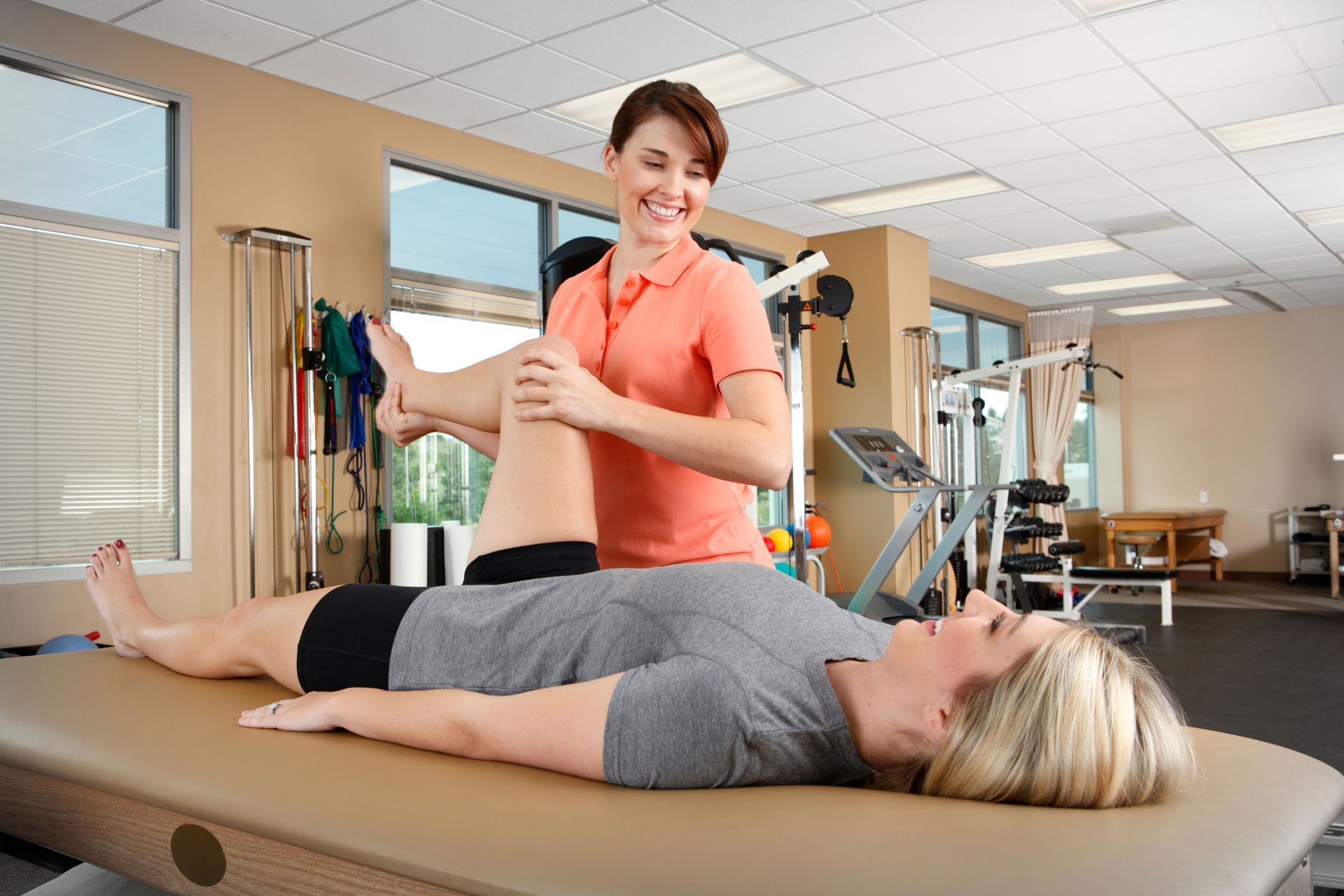
Neuromuscular control deficits significantly contribute to exacerbating kinetic chain dysfunction during rehabilitation from orthopedic injuries by impairing the coordination and timing of muscular responses essential for maintaining stability and proper movement mechanics. These deficits can lead to altered proprioception, resulting in diminished joint awareness and an increased risk of compensatory movement patterns that further stress interconnected muscle groups, ligaments, and joints within the kinetic chain. As a consequence, individuals may experience impaired force production, decreased neuromuscular efficiency, and heightened susceptibility to re-injury due to inadequate stabilization strategies. Furthermore, lack of adequate motor learning processes can hinder adaptive changes necessary for restoring functional movements post-injury; this not only prolongs recovery time but also complicates the rehabilitation process through maladaptive compensation pathways that reinforce dysfunctional biomechanics throughout various phases of healing. Hence, addressing neuromuscular control is vital for optimizing therapeutic outcomes and ensuring effective restoration of dynamic equilibrium across affected musculoskeletal systems during recovery from injury.

Exploring the Role of Kinetic Chain Dysfunction in Orthopedic Injuries

Exploring the Role of Kinetic Chain Dysfunction in Orthopedic Injuries

Contact Us

Alpine Physical Therapy and Fitness

  • Address: 3130 Alpine Rd Suite 370, Portola Valley, CA 94028
  • Phone: (650) 242-0777
  • Email: alpineptfit@mail.com

© Copyright - All Rights Reserved